A brain operation begins well before admission: you need to know whether symptoms require urgent assessment, what your scans show, which treatments are available and what preparation the hospital expects. By the end, you will understand the usual path from consultation and testing through surgery, monitoring, discharge and follow-up, including questions to ask before consenting.
Key takeaways
- Seek urgent assessment for weakness, facial drooping, seizures or sudden severe headache.
- Bring symptom timelines, medication details and previous scan reports to consultation.
- Doctors weigh diagnosis, symptoms, risks and alternatives before recommending brain surgery.
- Ask how hospital recovery, pathology results and follow-up appointments will be handled.
When to Seek Urgent Assessment and How to Prepare for Consultation
New weakness, facial drooping, difficulty speaking or understanding, a seizure, confusion, increasing drowsiness, repeated vomiting or a sudden severe headache needs urgent medical assessment. Do not wait for a routine brain operation consultation in Matunga when these symptoms appear.
1. For a planned consultation, bring MRI or CT images on the original disc or through the imaging portal. Carry the radiology reports, a complete medication list with doses, allergy history, previous operative notes and pathology reports.
2. Write a symptom timeline. Record when each symptom began, whether it is worsening, and any seizure, fall or change in vision, memory, speech or movement. This helps the neurosurgeon compare your account with the scan and examination.
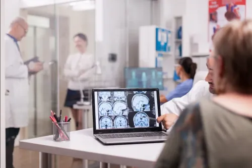
3. Ask directly whether the neurosurgeon has reviewed the actual MRI or CT images, not only the written report. A report can miss a detail that changes the proposed approach.
4. Use the consultation to establish the working diagnosis and the purpose of treatment. Ask about the expected benefit, major risks, alternatives such as observation, medication, biopsy or radiation, and what could happen without treatment.
Bring someone to take notes if the discussion is complex. If symptoms worsen while you are waiting, seek urgent assessment rather than rescheduling a routine appointment.
How Doctors Decide Whether Surgery Is Suitable
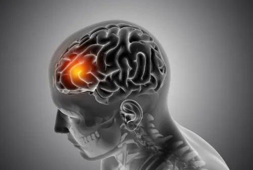
Surgery is suitable when its expected benefit outweighs the risks of the operation and of leaving the condition untreated. A scan alone cannot make that decision.
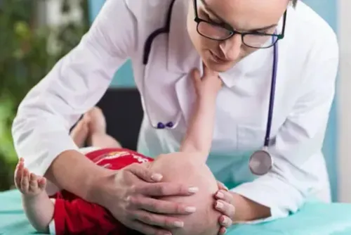
1. Your neurological examination checks strength, sensation, balance, coordination, vision, speech, memory and alertness. Doctors compare these findings with the lesion’s location, size, evidence of growth and the symptoms it causes.
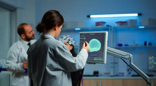
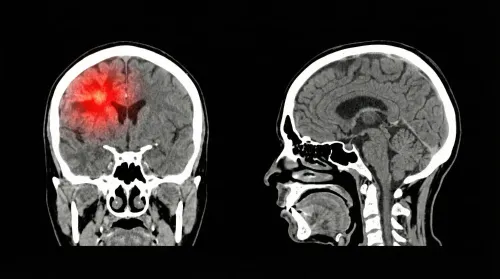
2. Contrast MRI can define tissue and areas of enhancement. CT shows bone, blood and urgent swelling. CT angiography maps nearby arteries; functional MRI and tractography help locate language, movement and vision pathways. Diffusion or perfusion imaging adds information about tissue behaviour or blood flow.
3. Blood tests check anaemia, clotting and kidney function before contrast or anaesthesia. An electrocardiogram and anaesthesia assessment identify medical problems that could change operative planning. Neuropsychological or seizure-related testing is added when memory, language or seizures matter.
4. The team compares observation, medication, biopsy, radiation, stereotactic radiosurgery and cranial surgery. It weighs likely benefit against bleeding, infection, seizures, stroke, swelling, anaesthetic complications and new neurological deficits. A small lesion causing no symptoms may need surveillance, while growth or worsening function can favour treatment.
A second opinion is valuable when the diagnosis is uncertain, the operation is high-risk or alternatives have not been explained. Ask the second neurosurgeon to review the original images and pathology, particularly before brain surgery in Matunga.
Which Operation and What Happens on the Day
The operation name tells you what the surgeon plans to access, remove or test; it does not mean every brain operation follows the same route.
| Procedure | What it means | When it applies |
|---|---|---|
| Craniotomy | A skull section is temporarily removed and replaced at closure. | Accessing a lesion or treating a condition beneath the bone. |
| Craniectomy | The bone is left off temporarily or permanently to create space for swelling. | Severe swelling makes immediate replacement unsafe. |
| Biopsy | A tissue sample is removed for diagnosis, not the whole lesion. | The diagnosis will determine further treatment. |
| Endoscopic surgery | A camera and narrow instruments pass through a small opening or natural corridor. | Selected conditions suited to minimally invasive access. |
| Skull-base approach | The route reaches the underside of the brain near nerves and blood vessels. | Lesions in the skull base region. |
| Awake craniotomy | You are sedated or asleep for selected stages, then name objects or move a limb while language or motor areas are mapped. | Lesions close to important functional areas; it is not routine. |
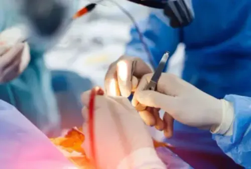
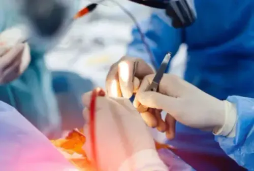
On admission, the team checks your consent, fasting status, allergies and medication instructions. Staff may prepare your hair, position and secure your head, and register image-guided navigation.
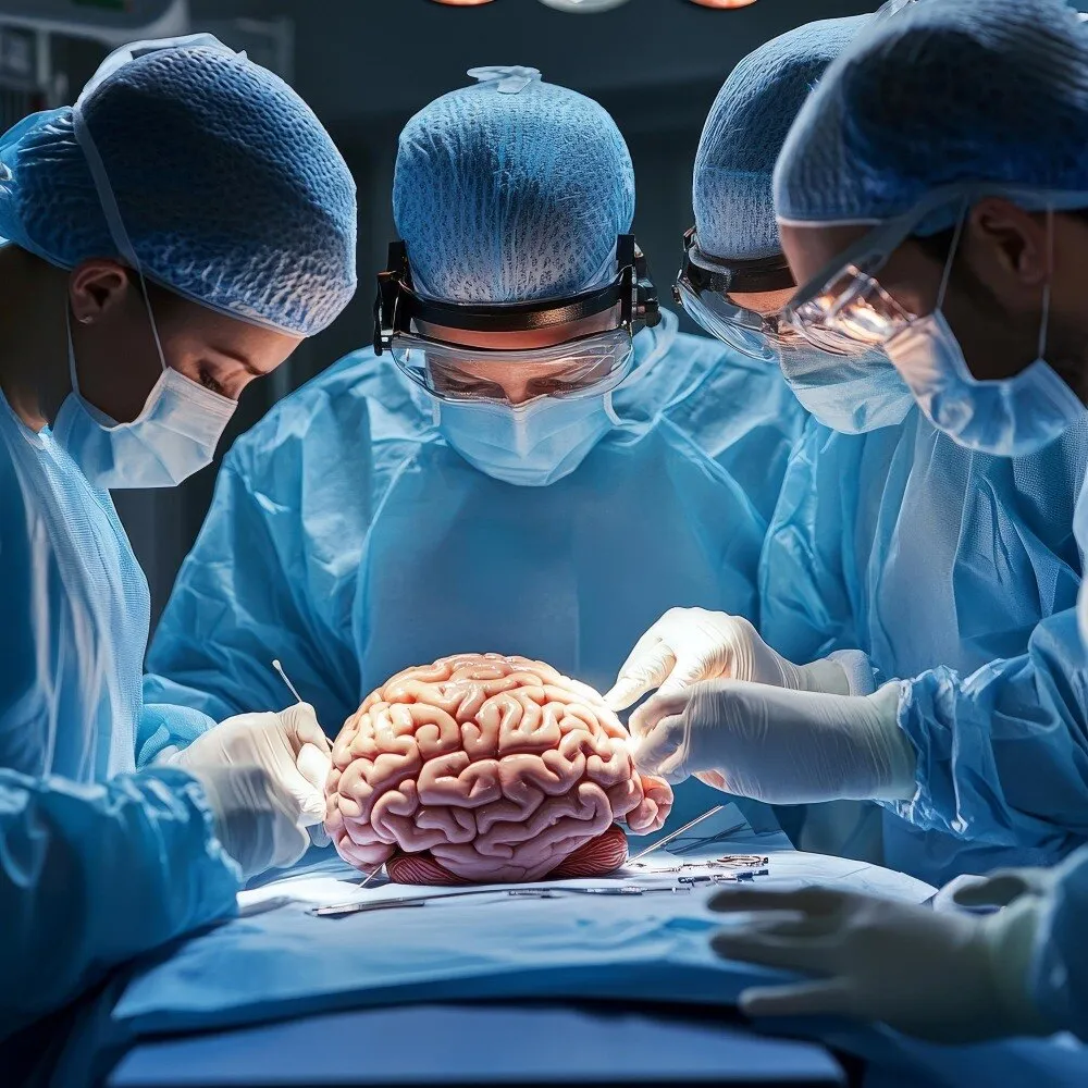
During the operation, monitoring of brain or nerve function can help protect critical pathways. The surgeon may collect a specimen for pathology. Before you leave the operating area, the team performs neurological checks; CT or MRI follows when the planned procedure or your condition requires it. Not every operation uses every step.
For anyone comparing brain cranial surgery in Matunga, confirm that the facility has neuroanaesthesia, intensive care, urgent CT or MRI, blood-bank support and rehabilitation pathways.
What Hospital Recovery Looks Like After Brain Surgery
After major cranial surgery, you usually spend time in a recovery area or neurocritical-care or intensive-care unit before moving to a ward when stable. Recovery does not follow a fixed timetable: an uncomplicated craniotomy may require several hospital days, while complications or rehabilitation needs can extend it.
1. Nurses and doctors repeatedly check consciousness, pupils, speech, strength, sensation, breathing and vital signs. They may order a postoperative CT or MRI to confirm the surgical result or investigate a new neurological change; imaging is not automatic after every operation.
2. On the ward, expect wound observation, pain and nausea treatment, medication review, mobilisation and checks for seizures or new neurological deficits. Weakness, speech or memory problems, seizures, swelling, infection or bleeding can prolong admission and lead to physiotherapy, speech therapy or other rehabilitation.
3. Headache, fatigue, scalp tenderness, numbness around the incision and temporary concentration or mood changes can continue after the wound heals. Arrange help with meals, transport, childcare and medicines before discharge; brain surgery recovery Matunga patients plan well is safer than assuming a rapid return to routine.
4. Do not set a return date for work, driving, exercise or strenuous activity without individual clearance. After discharge, seek urgent assessment for worsening weakness, facial drooping, speech difficulty, seizure, increasing drowsiness, severe headache or repeated vomiting.
Follow-Up, Pathology and Choosing the Right Hospital Support
After discharge, follow-up should review the wound, neurological symptoms, medicines, scan findings and rehabilitation needs. Tissue sent for histopathology can mean you leave before the final diagnosis is available. A tumour report may state its type, grade and molecular findings, guiding radiotherapy, chemotherapy, targeted treatment, seizure treatment or surveillance.
Seek urgent assessment rather than waiting for routine follow-up for:
- New or worsening weakness, facial drooping, speech difficulty or sudden vision change
- A seizure, increasing drowsiness, severe or rapidly worsening headache, or repeated vomiting
- Fever with wound redness or drainage, or clear fluid from the wound or nose
If you are researching brain surgery in Matunga what to expect, judge the complete care pathway, not the consultation room alone. Ask whether the proposed facility provides:
| Facility support | Why it matters |
|---|---|
| Round-the-clock neurosurgical cover and neuroanaesthesia | Prompt specialist decisions and safe anaesthesia |
| Neurocritical care, urgent CT or MRI and operating-room backup | Rapid response to deterioration or complications |
| Blood-bank support | Access to blood products if bleeding occurs |
| Rehabilitation or referral pathways | Continued help with weakness, speech, memory or mobility |
Dr. Anshu Warade can provide specialist assessment, but hospital capacity shapes brain surgery recovery in Matunga. Before admission, confirm who reviews the pathology report, who handles after-hours concerns and where rehabilitation will take place.
Related services
Brain (Cranial) Surgeries 🧠 Brain (Cranial) Surgeries by Dr. Anshu Warade Brain (Cranial) Surgeries involve advanced surgical procedures to treat conditions affecting the... View service → |
Neurological Examination 🧠 Neurological Examination by Dr. Anshu Warade A Neurological Examination is a detailed clinical assessment used to evaluate the functioning of the... View service → |
Frequently asked questions
When should I seek urgent assessment instead of waiting for a consultation?
Seek urgent medical assessment for new weakness, facial drooping, speech or comprehension difficulty, a seizure, confusion, increasing drowsiness, repeated vomiting or a sudden severe headache.
How do doctors decide whether brain surgery is suitable?
Doctors assess your diagnosis, symptoms, examination findings and scan results, then weigh the expected benefit against surgical risks and available alternatives.
What happens on the day of brain or cranial surgery?
The surgical team confirms the planned operation, completes preoperative checks and explains what to expect before anaesthesia, the procedure and immediate monitoring.
What does hospital recovery involve after brain surgery?
Recovery includes monitoring neurological function, managing pain and other symptoms, checking progress and planning discharge instructions, follow-up and further care.
Why are pathology and follow-up important after brain surgery?
Pathology can identify the nature of tissue removed during surgery, while follow-up helps review results, recovery and any additional treatment or monitoring needed.
Related services
Skull Base Neurosurgery 🧠 Skull Base Neurosurgery by Dr. Anshu Warade Skull Base Neurosurgery is a highly specialized field that focuses on treating complex tumors and... View service → |
Pediatric Neurosurgery 🧠 Pediatric Neurosurgery by Dr. Anshu Warade Pediatric Neurosurgery focuses on diagnosing and treating brain, spine, and nerve conditions in... View service → |
Endoscopic Brain Surgery 🧠 Endoscopic Brain Surgery by Dr. Anshu Warade Endoscopic Brain Surgery is an advanced, minimally invasive neurosurgical technique that uses a thin... View service → |
Keywords