A stroke is not automatically treated with an operation: a blocked artery, bleeding, swelling, hydrocephalus and an aneurysm require different decisions. By the end, you will know which interventions may protect brain tissue, when emergency transfer matters, and what questions to ask about treatment and recovery in the Dadar area.
Key takeaways
- Call emergency services; do not drive to an outpatient appointment.
- CT identifies bleeding before angiography maps blocked or ruptured vessels.
- Clot removal works by reopening a blocked artery through a catheter.
- Ask how swelling, bleeding, aneurysms and rehabilitation will be managed.
Which stroke needs urgent surgery or a catheter procedure?
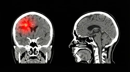
Call emergency services immediately; do not drive to an outpatient neurosurgical appointment. Doctors first use a non-contrast CT scan to identify bleeding, then CT angiography to locate a blocked or ruptured vessel. Treatment depends on that result, not simply on the symptoms or the patient’s location.
| Type | What the scan suggests | Urgent treatment considered |
|---|---|---|
| Ischaemic stroke | A blocked artery, with no initial bleed; CT angiography may show a large-vessel occlusion | Intravenous thrombolysis for eligible patients and mechanical thrombectomy through a catheter, usually within 6 hours and up to 16–24 hours for selected imaging profiles |
| Haemorrhagic stroke | Blood inside the brain or around it; CT shows volume, location, pressure, hydrocephalus or midline shift | Blood-pressure control, haematoma evacuation, drainage of hydrocephalus, decompressive craniectomy, or catheter treatment for an aneurysm |
Any search for stroke related surgical interventions benefits in Dadar must not delay transfer to a hospital with imaging and emergency stroke capability. A clot-removal procedure suits a removable large-artery clot, not every ischaemic stroke; open surgery suits dangerous swelling or selected bleeds.
- Note the exact time the person was last known well.
- Keep the person lying safely, and give no food, drink, aspirin or other medicine unless emergency clinicians instruct you.
- Take the medication list, anticoagulant name and dose, medical history, and identification to hospital.
- Ask whether CT angiography, thrombectomy, neurosurgery and intensive care are available immediately.
How clot-removal procedures restore blood flow
For families searching stroke surgery benefits Dadar, the key question is whether blood flow can be restored before brain tissue becomes permanently damaged. A non-contrast CT rules out bleeding, while CT angiography identifies a blocked artery; doctors also assess the clot location, infarct size, previous disability and time last known well.
- Intravenous thrombolysis: An eligible patient may receive clot-dissolving medicine, usually within 4.5 hours of symptom onset. It is not suitable for every patient, especially when bleeding risk or established large infarction is present.
- Mechanical thrombectomy: A catheter removes a clot from a major artery, often through the groin or wrist, without opening the skull. Selected patients benefit within 6 hours, while favourable perfusion scans and clinical findings can support treatment 6 to 16 or 24 hours after the patient was last known well.
- Carotid endarterectomy or stenting: These procedures reduce the risk of another stroke when severe extracranial carotid narrowing caused the symptoms. They do not repair brain tissue already injured by the infarct; timing depends on neurological stability, infarct size, stenosis and operative risk.
Call emergency services rather than travelling to an outpatient appointment. A hospital without immediate thrombectomy capability should arrange rapid transfer. Restoring flow does not replace recovery care: physiotherapy, speech-language therapy, swallowing assessment, occupational therapy and cognitive rehabilitation address the resulting disabilities, and recovery varies widely.
How surgery controls swelling, pressure and brain bleeding
A large infarct can swell enough to compress the brainstem or push brain tissue across the midline. A haemorrhage can do the same through expanding blood, hydrocephalus, or rising intracranial pressure. Non-contrast CT, CT angiography, neurological deterioration, bleed location and overall medical condition guide the decision; surgery is not routine treatment for every stroke.
| Operation | Benefit | Main risks and limits |
|---|---|---|
| Decompressive hemicraniectomy | Removes part of the skull, usually for life-threatening swelling after a large middle-cerebral-artery infarct; within about 48 hours in selected patients, it reduces mortality. | Survival can mean major disability; risks include infection, bleeding, seizures and later reconstruction surgery. |
| Posterior-fossa decompression | Creates space when a swollen cerebellar infarct compresses the brainstem or blocks cerebrospinal-fluid flow. | Anaesthesia, bleeding and neurological injury remain possible; timing is critical. |
| Haematoma evacuation | Removes selected brain bleeds that cause deterioration, substantial pressure, or dangerous posterior-fossa compression. | The operation can injure healthy tissue, rebleeding can occur, and it cannot reverse damage already caused. |
| External ventricular drain | Drains cerebrospinal fluid quickly when intraventricular blood or swelling causes hydrocephalus or raised pressure. | It is temporary, not a cure; infection, blockage, bleeding and over-drainage can occur. |
Mechanical thrombectomy removes a large-artery clot through a catheter and often avoids open brain surgery. It does not treat swelling already established. Families researching brain surgery after stroke Dadar should ask whether the centre can provide urgent imaging, neurosurgical review and intensive care. Pressure control still needs physiotherapy, speech-language therapy, swallowing assessment and other rehabilitation.
When an aneurysm or other vascular cause needs planned treatment
Clipping, coiling, flow diversion, and treatment of an arteriovenous malformation (AVM) or cavernoma target an identified blood-vessel abnormality; emergency stroke surgery treats immediate damage such as swelling, pressure, hydrocephalus, or a dangerous haematoma. A ruptured aneurysm still needs urgent assessment and often early securing, not a routine outpatient appointment.
| Option | What it does | When it applies |
|---|---|---|
| Clipping | Places a clip across the aneurysm neck during open surgery | A durable option when the aneurysm’s shape or location suits microsurgery |
| Coiling | Packs the aneurysm from inside with detachable coils delivered by catheter | A less invasive option for selected aneurysms, including some ruptured aneurysms |
| Flow diversion | Uses a stent-like mesh to redirect blood away from the aneurysm | Selected wide-neck or complex aneurysms; it requires antiplatelet medication |
| AVM treatment | Removes the malformation, closes it with catheter embolisation, or targets it with focused radiation | Choice depends on size, location, bleeding history, and surgical risk |
| Cavernoma treatment | Removes the compact cluster of abnormal vessels | Considered after bleeding, seizures, or progressive neurological symptoms when access is reasonable |
Unlike decompressive craniectomy or haematoma evacuation, these procedures do not reverse established brain injury. Stroke surgery benefits dadar claims mean little without CT, CT angiography, MRI, or catheter angiography showing a treatable lesion, plus a plan for rehabilitation and follow-up.
How to choose a Dadar-area stroke pathway and plan recovery
Choose a Dadar-area pathway by its emergency capability and transfer plan—not by proximity or the label “stroke neurosurgeon Dadar.” Confirm that the receiving hospital can perform immediate non-contrast CT, CT angiography, stroke-neurology and neurosurgical assessment, endovascular treatment, intensive-care monitoring, and rapid transfer.
If thrombectomy is unavailable, delay while seeking a local appointment can cost brain function.
Ask the team to link the proposed treatment to your scan, vessel involved, infarct size, bleeding risk, previous disability and medical conditions. Request the named operator’s experience, audited outcomes and a plain explanation of what happens if the procedure fails.
- For thrombectomy, ask about intracranial bleeding, vessel injury, clot migration, contrast reaction and groin or wrist bleeding.
- For decompressive craniectomy, ask about survival with disability, infection, seizures, repeat surgery and the timing of skull reconstruction.
- Before discharge, confirm physiotherapy, occupational therapy, speech-language therapy, swallowing assessment, cognitive rehabilitation and prevention of pressure injuries, clots and another stroke.
| Option | What to verify | Why it matters |
|---|---|---|
| Emergency pathway | CT/CTA, ICU, specialist cover and transfer protocol | Treatment delays worsen eligibility and outcome |
| Procedure choice | Clot, swelling, haemorrhage or hydrocephalus on imaging | Different problems require different operations |
| Recovery team | Named rehabilitation services and review dates | Surgery treats the crisis, not every resulting disability |
Dr. Anshu Warade can help clarify neurosurgical risks and recovery goals, but emergency assessment must come through a stroke-capable hospital.
Frequently asked questions
Which strokes need urgent surgery or a catheter procedure?
Doctors use a non-contrast CT scan to identify bleeding, followed by CT angiography to locate a blocked or ruptured vessel. A blocked large artery may require catheter-based clot removal, while bleeding, swelling or pressure may require surgery.
How do clot-removal procedures restore blood flow?
A specialist guides a catheter through the blood vessels to the blocked artery and removes the clot, restoring blood flow to threatened brain tissue.
When is surgery used to control swelling, pressure or brain bleeding?
Surgery is considered when bleeding or swelling raises pressure inside the skull and threatens brain function. Treatment depends on the scan findings, the bleed’s location and the patient’s condition.
When does an aneurysm or other vascular cause need planned treatment?
After the emergency phase, doctors may plan treatment for an aneurysm or another vessel abnormality identified on vascular imaging. The team compares options such as endovascular treatment and open surgery.
How do you choose a Dadar-area stroke pathway and plan recovery?
Choose an emergency-capable hospital with rapid CT, CT angiography, stroke specialists, neurosurgical support and rehabilitation coordination. Ask who will manage follow-up, medicines, swallowing, movement, speech and return-home planning.
Keywords