A suspected severe head injury in Santacruz needs emergency stabilisation and access to non-contrast CT, neurosurgical assessment, an operating theatre and intensive-care support—not a routine clinic appointment. By the end, you will understand what CT findings drive surgery, how the main operations differ, and when close ICU observation is safer than immediate intervention.
Key takeaways
- Call 112, keep the neck still, and avoid moving the injured person.
- An epidural or subdural haematoma with brain pressure can require urgent surgery.
- Burr-hole drainage, craniotomy and fracture repair treat different injury patterns.
- A normal first CT does not rule out deterioration or a later bleed.
What happens after a serious head injury in Santacruz?
Call 112 in India or the local ambulance service immediately. Keep the injured person still, support the head, and do not move the neck unless staying there creates immediate danger. Brain injury surgery near Santacruz is an emergency hospital pathway, not a routine neighbourhood consultation.
- Paramedics protect the airway and cervical spine, assess breathing and oxygenation, support circulation, and prevent further movement before transport.
- They record the Glasgow Coma Scale, pupil size and reaction, limb movement, vomiting, seizures, and any loss of consciousness.
- Emergency-department triage identifies patients needing immediate resuscitation, a non-contrast CT scan of the head, and cervical-spine assessment.
- The receiving hospital must provide neurosurgical review, anaesthesia, an operating theatre, and neurocritical-care or ICU monitoring.
Severe TBI can worsen during transport, so do not wait for an outpatient appointment. Carry the time and mechanism of injury, the patient’s anticoagulant or antiplatelet medicines, seizures, vomiting, allergies, and baseline neurological function.
Ask whether the hospital can manage traumatic brain injury TBI surgery Santacruz emergencies on site or arrange rapid transfer; a clinic without CT, neurosurgery and ICU access cannot safely replace that pathway.
Which CT findings make surgery urgent, and when is observation reasonable?
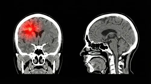
Surgery becomes urgent when a clot or swollen area is compressing the brain, consciousness is falling, or pupils change. An abnormal CT alone does not mandate an operation; traumatic brain injury surgery details depend on size, location, midline shift, neurological examination and intracranial pressure.
| Finding | Surgical concern | When observation may be reasonable |
|---|---|---|
| Acute epidural haematoma | Evacuate when volume exceeds 30 cm³ | Neurosurgical-centre observation when under 15 mm thick, under 5 mm shift, GCS above 8 and no focal deficit |
| Acute subdural haematoma | Evacuate when thickness exceeds 10 mm or shift exceeds 5 mm | A smaller clot needs close review if GCS drops by 2 points, pupils become unequal or fixed and dilated, or pressure exceeds 20 mmHg in coma |
| Contusion or intraparenchymal haematoma | Frontal or temporal lesion over 20 cm³ with at least 5 mm shift, or any lesion over 50 cm³, may require surgery in a deteriorating patient | Observe only with serial examinations and imaging when mass effect is controlled |
| Posterior-fossa lesion | Urgent treatment if it compresses the fourth ventricle, causes hydrocephalus or produces neurological decline | Observation is unsafe when brainstem or cerebrospinal-fluid pathways are threatened |
| Traumatic subarachnoid haemorrhage | Decision follows cistern compression, mass effect and neurological decline | A small haemorrhage without compression can be monitored |
An abnormal CT with a post-resuscitation GCS of 3–8 can require intracranial-pressure monitoring; pressure above 22 mmHg is treated. A search for tbi surgery santacruz should therefore focus on a hospital able to repeat CT, monitor pupils and consciousness, provide ICU care and operate quickly—not on the scan label alone.
How do burr-hole drainage, craniotomy and skull-fracture repair differ?
The operation depends on whether the problem is a drainable blood collection, damaged skull, injured brain tissue or dangerous swelling. These traumatic brain injury surgery details explain why “tbi surgery santacruz” can involve different procedures.
| Option | What it means | When it applies |
|---|---|---|
| Burr-hole drainage | One or more small skull openings release a suitable blood collection; a drain remains when needed. | Selected collections, often when a smaller access route is sufficient. |
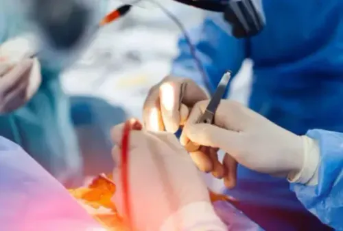
| Craniotomy | A larger scalp incision creates a temporary bone flap. The surgeon opens the dura, removes clot or damaged tissue, controls bleeding, manages the dura, checks brain relaxation, places drains if required and replaces the flap with plates or sutures. | A mass lesion or structural damage requiring direct treatment. |
| Decompressive craniectomy | A skull segment is removed and deliberately not replaced. | Severe swelling or refractory pressure that needs room for the brain. |
| Fracture repair | Depressed fragments are lifted away from the brain; damaged dura is repaired when necessary. | A depressed skull fracture or open skull injury. |
| Open-injury repair | Contaminated tissue is cleaned, bleeding is controlled, the dura is repaired and the scalp is closed. | An open wound, with infection prevention tailored to contamination. |
The practical sequence usually includes:
- General anaesthesia, hair and skin preparation, and head positioning in a fixed support.
- Monitoring, treatment of bleeding and the selected repair.
- Postoperative ventilation, repeat imaging or ICU care when brain swelling or reduced consciousness requires it.
Removing a clot treats a mass lesion; ventilation, blood-pressure support, sedation and pressure control treat secondary brain injury, whether surgery occurs or not.
Why is part of the skull left out, and what risks must families compare?
Leaving the bone flap out is not routine clot removal. A decompressive craniectomy creates space when brain swelling or refractory intracranial pressure threatens compression despite medical treatment; a craniotomy usually replaces the bone after the clot or damaged tissue is treated.
| Procedure | Purpose | What happens to the bone |
|---|---|---|
| Craniotomy | Remove a clot or treat structural damage | Replaced during the same operation |
| Decompressive craniectomy | Relieve dangerous swelling or pressure | Left out temporarily |
| Cranioplasty | Reconstruct the skull | Replaces the flap or a custom implant later |
The removed bone may be stored in a specialised freezer or placed in an abdominal tissue pocket, depending on hospital practice. Ask which method the hospital uses. After swelling settles and the patient is medically ready, a separate cranioplasty restores skull protection; it is reconstruction after the emergency, not the decompression itself.
Surgery can lower mortality in refractory intracranial hypertension while leaving some survivors with severe disability. Compare the intended benefit with rebleeding, infection, seizures, stroke, worsening swelling, cerebrospinal-fluid leak, anaesthesia complications, blood clots and persistent neurological disability. Discuss likely function—speech, movement, memory and independence—not survival alone.
For traumatic brain injury surgery details, ask:
- Is the goal clot removal, fracture repair, pressure control, or several goals?
- What happens without surgery, and is a second operation likely?
- When could cranioplasty be considered?
These questions matter when assessing traumatic brain injury TBI surgery Santacruz.
What if the first CT does not show an immediate need for surgery?
A stable first CT does not mean “nothing needs doing.” Non-operative care is active treatment: doctors may admit you for repeated neurological observations, pupil checks, blood-pressure and oxygen control, seizure treatment and repeat CT when symptoms change or a lesion could expand.
| Option | What it involves | When it applies |
|---|---|---|
| Monitored observation | Serial examinations and repeat CT | Stable lesion, stable pupils and consciousness |
| Neurocritical care | Ventilation, sedation, head elevation, blood-pressure support and pressure monitoring | Severe TBI with GCS 3–8 after resuscitation and an abnormal CT |
| Operation | Clot evacuation, fracture repair or decompression | Neurological decline, dangerous mass effect or uncontrolled pressure |
For severe TBI, intracranial pressure above 22 mmHg is treated; cerebral perfusion is commonly targeted around 60–70 mmHg. Fever and seizures also need treatment. Routine corticosteroids, including high-dose methylprednisolone, are not treatment for severe TBI.
Antiseizure medicine may be prescribed during the first 7 days in selected severe injuries, but it does not automatically continue indefinitely.
Before choosing care for traumatic brain injury tbi surgery santacruz or brain injury surgery near santacruz, confirm access to CT, neurosurgery, an operating theatre and ICU. If you contact Dr. Anshu Warade, ask where emergency assessment and neurocritical care will occur; a routine consultation must not delay ambulance transfer.
Return urgently for:
- Worsening drowsiness, repeated vomiting, new weakness, unequal pupils, seizure, severe headache or confusion.
Related service
Brain (Cranial) Surgeries 🧠 Brain (Cranial) Surgeries by Dr. Anshu Warade Brain (Cranial) Surgeries involve advanced surgical procedures to treat conditions affecting the... View service → |
Frequently asked questions
What happens after a serious head injury in Santacruz?
Call 112 or the local ambulance service, keep the person still, support the head and avoid moving the neck. Emergency staff assess consciousness, pupils, movement and breathing before arranging CT imaging and neurosurgical care.
Which CT findings make traumatic brain injury surgery urgent?
A large epidural or subdural haematoma, significant midline shift, compressed brain structures, severe swelling or a depressed skull fracture can make surgery urgent. The decision also depends on consciousness, pupil changes and neurological deterioration.
When is observation reasonable after a head injury?
Observation can be appropriate when the bleed is small, the neurological examination is stable and repeat examinations and CT scans are available. New drowsiness, vomiting, weakness, seizures or unequal pupils require immediate reassessment.
How do burr-hole drainage, craniotomy and skull-fracture repair differ?
Burr-hole drainage uses a small opening to remove selected blood collections. A craniotomy temporarily removes a larger bone flap to evacuate a clot and control bleeding. Skull-fracture repair addresses depressed or open fractures and damaged tissue.
Why is part of the skull left out after brain injury surgery?
A decompressive craniectomy leaves the bone flap out when brain swelling needs extra space. The opening is protected while swelling settles, and a later cranioplasty replaces the bone or uses an implant.
Keywords