Numbness, weakness, pain, imbalance and loss of coordination can arise from a peripheral nerve, nerve root, plexus or brain, so the correct operation cannot be chosen from the incision size or location alone. You will be able to identify the likely anatomical problem, understand how testing guides the choice, compare surgical and non-surgical options, and prepare for a safer consultation in Matunga.
Key takeaways
- Peripheral nerve surgery treats nerves outside the brain and spinal cord.
- MRI, nerve conduction studies and examination help locate the problem.
- Ask about anesthesia, hospital stay, recovery milestones and follow-up.
- Choose surgery after diagnosis, progression and alternatives have been reviewed.
Peripheral nerve surgery and endoscopic brain surgery treat different anatomical problems
Peripheral nerve surgery treats a nerve outside the brain and spinal cord. It covers carpal tunnel syndrome, cubital tunnel syndrome, traumatic nerve laceration, focal compression, selected nerve tumors and brachial plexus injury. It is not spinal surgery, brain surgery or treatment for diffuse neuropathy caused by diabetes, vitamin deficiency or systemic disease.
The operation depends on the damage:
- Decompression releases a tight tunnel around a nerve.
- Neurolysis frees a nerve trapped in scar tissue.
- Direct repair joins freshly cut nerve ends.
- Nerve grafting bridges a gap with donor nerve.
- Nerve transfer connects a working donor nerve to a damaged target.
Endoscopic brain surgery is an access method for a defined intracranial problem, not a diagnosis. An endoscopic third ventriculostomy creates a cerebrospinal-fluid pathway for selected obstructive hydrocephalus. An endoscopic endonasal route reaches selected skull-base lesions through the nose. Neither is automatically a smaller or safer substitute for every craniotomy.
Searches for “peripheral nerve surgery Matunga” and “endoscopic brain surgery Matunga” describe location, not suitability. This peripheral nerve surgery comparison depends on the diagnosis, anatomy, imaging, electrodiagnostic findings and specialist expertise—not the neighbourhood or incision size.
Symptoms and tests reveal whether the problem is in a nerve or the brain
Numbness or weakness confined to one hand, one named nerve, or an area injured in an accident points more toward a peripheral problem. Tingling triggered by wrist or elbow compression fits entrapment; imbalance, seizures, sudden severe headache, visual loss, reduced consciousness, or weakness on one side of the body raises concern for the brain.
Rapidly progressive weakness or new bladder or bowel loss needs prompt assessment.
| Pattern | More suggestive of | Why it matters |
|---|---|---|
| Hand weakness or numbness | Peripheral nerve | May match median, ulnar, or another named nerve |
| Symptoms after laceration or limb injury | Peripheral nerve or plexus | The injury may identify the damaged level |
| Imbalance, seizure, visual loss, or reduced consciousness | Brain | These symptoms require urgent neurological assessment |
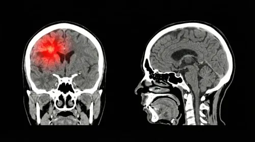
A focused neurological examination tests strength, sensation, reflexes, coordination, gait, cranial nerves, and muscle wasting, helping localize the problem before testing. MRI can show brain lesions, compression, masses, or denervation-related muscle change. MR neurography follows a peripheral nerve or plexus. CT adds detail about bone, acute bleeding, and surgical anatomy.
Electromyography and nerve-conduction studies localize dysfunction and estimate severity, but an equivocal result does not exclude early focal entrapment. No test replaces examination. This distinction should guide any peripheral nerve surgery comparison or search for nerve surgery options Matunga.
The access route, anesthesia and hospital course are not interchangeable
Anatomy determines the operation: peripheral nerve surgery reaches a nerve outside the brain and spinal cord, while endoscopic brain surgery enters the skull or a skull-base corridor. A peripheral procedure may use an incision near the wrist, elbow, limb, neck or injury site. Decompression or neurolysis is different from direct repair, grafting or nerve transfer.
| Option | Access and target | Imaging and expected course |
|---|---|---|
| Peripheral nerve surgery | Exposed nerve at a compression or injury site; limb or neck incision | MRI, MR neurography or electrodiagnostic findings guide treatment; anesthesia, operating time and admission vary |
| Endoscopic ventricular surgery | Small cranial opening into the ventricles | Neuronavigation matches instruments to MRI or CT; general anesthesia and neurological monitoring may be required |
| Endoscopic endonasal surgery | Nose-to-skull-base route for selected lesions | MRI or CT defines the corridor; CSF leak, visual injury and nasal complications remain possible |
An endoscopic third ventriculostomy makes a cerebrospinal-fluid pathway for selected obstructive hydrocephalus, but it does not suit every hydrocephalus pattern and can close later. Endonasal access is not an automatic substitute for craniotomy.
A search for endoscopic brain surgery matunga or peripheral nerve surgery compared with endoscopic brain surgery matunga describes location, not one standard operation. Ask the treating team for procedure-specific anesthesia, operating-time and hospital-stay figures.
Surgery is chosen after diagnosis, progression and failed alternatives are weighed
Peripheral decompression is considered when symptoms localize to a compressible point and splinting, physiotherapy, pain treatment or a clinically appropriate steroid injection has failed. Progressive weakness, muscle wasting or documented nerve damage makes delay more harmful.
Traumatic laceration and severe brachial-plexus injury need different timing, often involving repair, grafting or transfer rather than simple decompression. For selected nerve tumors, surveillance may be safer than immediate removal. Long-standing axonal loss cannot be reversed simply by releasing pressure.
The peripheral nerve surgery comparison with endoscopic brain surgery in Matunga should include procedure-specific risks, not incision size:
| Option | Main risks to compare |
|---|---|
| Peripheral decompression or repair | Persistent pain or numbness, scar sensitivity, neuroma, infection, incomplete recovery and injury to nearby nerves |
| Endoscopic brain surgery | Bleeding, infection, seizures, neurological or visual deficits, CSF-flow failure and CSF leakage requiring repair or lumbar drainage |
Before irreversible surgery, ask for:
- The surgeon’s own complication and incomplete-recovery rates for the exact operation, not a hospital-wide average.
- An explanation when imaging does not match your symptoms; a scan abnormality alone does not prove the pain source.
- Alternatives such as observation, continued therapy or tumor surveillance, plus a second opinion when the diagnosis, timing or expected benefit remains uncertain.
Recovery depends on decompression, repair or brain access—not incision size alone
Decompression removes pressure, but it does not instantly restore an injured nerve. Strength and sensation can return gradually, especially when compression has lasted a long time. Direct nerve repair, grafting and nerve transfer require a longer wait: after an initial delay, repaired or grafted axons commonly regenerate at about 1 millimetre per day.
Recovery can take months to years and remain incomplete.
Hand therapy, physiotherapy, wound care, lifting limits, driving and return-to-work timing must match the exact operation. A small incision does not guarantee a short recovery. A peripheral nerve surgery Matunga plan and an endoscopic brain surgery Matunga plan should therefore specify the procedure, milestones and follow-up rather than rely on incision size.
Endoscopic brain recovery depends on the route and diagnosis. Follow-up may assess neurological function and cerebrospinal-fluid flow, with nasal care after an endonasal route or seizure precautions when relevant.
Seek urgent medical advice instead of waiting for routine review if you develop:
- New weakness or worsening balance
- A seizure, severe headache, fever or reduced consciousness
- Clear fluid draining from the nose or wound
These symptoms can signal bleeding, infection, CSF leakage or neurological deterioration.
A Matunga consultation should confirm expertise, logistics and the complete follow-up plan
Matunga does not determine suitability: peripheral nerve surgery compared with endoscopic brain surgery in Matunga is a location search, not a treatment choice. Refer elsewhere if the required peripheral-nerve subspecialty, endoscopic route, imaging, pathology or rehabilitation service is unavailable nearby. That is sensible planning for nerve surgery options Matunga, not a setback.
| Check | Ask | Why it matters |
|---|---|---|
| Expertise | Does the surgeon regularly perform the exact decompression, repair, graft, transfer or endoscopic route proposed? | Techniques and outcomes differ. |
| Logistics | Where will MRI, MR neurography, CT, EMG, nerve-conduction studies, surgery, pathology and postoperative therapy occur? | Separate facilities can delay decisions or rehabilitation. |
| Continuity | How does emergency access work after discharge, and is follow-up convenient? | New weakness, fever or other urgent problems need rapid review. |
Bring imaging discs and reports, EMG and nerve-conduction reports, medication and anticoagulant lists, and previous operative records.
Ask for the exact diagnosis, surgical goal, alternatives including observation, expected functional outcome, surgeon-specific complication experience, hospital stay, complete follow-up plan and immediate-contact warning signs. Dr. Anshu Warade can be considered for matching these findings and plans to an appropriate neurosurgical pathway.
Related services
Endoscopic Brain Surgery 🧠 Endoscopic Brain Surgery by Dr. Anshu Warade Endoscopic Brain Surgery is an advanced, minimally invasive neurosurgical technique that uses a thin... View service → |
Neurological Examination 🧠 Neurological Examination by Dr. Anshu Warade A Neurological Examination is a detailed clinical assessment used to evaluate the functioning of the... View service → |
Frequently asked questions
What is the difference between peripheral nerve surgery and endoscopic brain surgery?
Peripheral nerve surgery treats nerves outside the brain and spinal cord, such as the median, ulnar or brachial plexus nerves. Endoscopic brain surgery reaches a brain or skull-base problem through a small corridor using an endoscope.
Which symptoms suggest a peripheral nerve problem?
Numbness, tingling, weakness or pain following a specific nerve distribution can suggest peripheral nerve compression or injury. Examination and tests are needed to distinguish this from spinal disease, brain disease or diffuse neuropathy.
What tests help determine whether surgery is needed?
A neurological examination, nerve conduction study, electromyography, ultrasound or MRI can help identify nerve compression, injury or a mass. Brain conditions may require MRI or CT with sequences selected for the suspected lesion.
Are the anesthesia and hospital stay the same for both operations?
No. The anesthesia, access route, monitoring, hospital stay and risks depend on the operation and the condition being treated. A surgeon should explain these details before consent.
When is peripheral nerve or endoscopic brain surgery considered?
Surgery is considered after confirming the diagnosis and weighing symptom progression, nerve or brain damage, non-surgical treatment and the expected benefit. A procedure is not automatically appropriate because an abnormal scan is present.
Related service
Pediatric Neurosurgery 🧠 Pediatric Neurosurgery by Dr. Anshu Warade Pediatric Neurosurgery focuses on diagnosing and treating brain, spine, and nerve conditions in... View service → |
Keywords