Endoscopic brain surgery is not one operation or one route: the surgeon may work through the nose, a small skull opening, or the brain’s fluid spaces. By the end, you will know how doctors select the route, what preparation and risks to expect, how recovery is monitored, and how to compare a surgeon’s qualifications without relying on the word “best.”
Key takeaways
- Review MRI or CT scans and discuss surgical routes before consenting.
- Ask whether an endoscopic approach suits your diagnosis and anatomy.
- Discuss vision, hormone, infection, bleeding and cerebrospinal-fluid risks.
- Confirm discharge instructions, warning signs and follow-up appointments.
What happens during endoscopic brain surgery, from consultation to discharge?
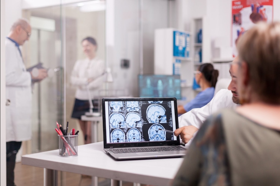
Endoscopic brain surgery explained begins with a consultation, neurological examination and review of MRI or CT scans. The surgeon discusses the diagnosis, route, alternatives and risks before written consent. An anaesthetist assesses you before general anaesthesia; after the operation, nurses monitor vision, strength, hormone-related symptoms, breathing and cerebrospinal-fluid drainage.
Discharge follows when your examination, pain control and recovery are stable, with specific wound, nasal-care and follow-up instructions. A search for endoscopic brain surgery bandra should not replace this diagnosis-specific assessment.
| Route | Access and purpose | Typical selected use |
|---|---|---|
| Endoscopic endonasal | Through the nasal cavity and sphenoid sinus to reach the skull base | Pituitary tumours and selected skull-base lesions |
| Endoscope-assisted keyhole | Through a small skull opening, using an endoscope for visualisation | Lesions reachable through a limited cranial corridor |
| Intraventricular endoscopy | Through a small opening into the brain’s fluid spaces | Hydrocephalus or intraventricular cysts |
- Endoscopic third ventriculostomy, or ETV, creates an opening in the floor of the third ventricle so cerebrospinal fluid bypasses an obstruction. It treats selected hydrocephalus patterns; it does not remove a tumour, and some patients later need a shunt or another procedure.
- The safest route depends on the lesion’s position, size, consistency, blood supply and relationship to nerves and vessels. A narrow corridor does not eliminate risks such as bleeding, infection, visual or hormonal injury, or cerebrospinal-fluid leakage.
- Before discharge, ask which symptoms require urgent review and when MRI, hormone tests or visual assessment will occur.
Which patients are suitable for an endoscopic approach?
An endoscopic approach suits a patient when the route reaches the target safely and allows adequate treatment. A generic tumour label, an online “best brain doctor” result or a hospital’s Bandra location cannot establish suitability.
The decision uses contrast MRI, CT when bone anatomy matters, tumour size and location, symptoms, age, previous treatment, blood-vessel and cranial-nerve involvement, and the available surgical corridor. For pituitary disease, hormone results and formal visual-field testing add important evidence; a scan alone cannot show the full treatment risk.
| Option | What it offers | When it may fit |
|---|---|---|
| Endoscopic surgery | A narrow route through the nose or a small skull opening | The target is reachable with adequate control and limited disruption |
| Open craniotomy or microscopic surgery | Wider exposure and stronger control of bleeding | The lesion is large, deeply positioned, vascular or close to critical nerves and vessels |
| Stereotactic treatment, radiosurgery, medication or observation | Treatment without a major operation | The lesion is small, slow-growing, inaccessible or causing no symptoms |
The choice is not simply between “minimally invasive brain surgery Bandra” and open surgery. A larger operation may be safer when the endoscopic corridor cannot expose the whole lesion or control bleeding. Planned endoscopic surgery can also require conversion or staged treatment if anatomy differs from the scans.
An endoscopic brain surgery explained consultation should cover these alternatives, including why observation or non-surgical treatment is unsuitable.
What does “minimally invasive” mean, and what risks should you discuss?
A smaller opening reduces tissue disruption; it does not make surgery risk-free. Before recommending it, the team commonly performs a neurological examination, contrast MRI, CT for bone detail, medication review and anaesthesia assessment. Pituitary disease also calls for endocrine testing, and formal visual-field testing may be needed.
The scans must show that a narrow corridor reaches the target and permits adequate treatment.
| Route | Access | Key consideration |
|---|---|---|
| Endonasal endoscopy | Nose and sphenoid sinus | Skull-base barrier reconstruction |
| Endoscope-assisted surgery | Small skull opening | Limited working angle |
| Intraventricular endoscopy | Small opening into the ventricle | Selected fluid-flow or cyst problems |
Discuss bleeding, vascular injury, infection, cerebrospinal-fluid leakage, meningitis, visual deterioration, pituitary hormone deficiency, diabetes insipidus, seizures and neurological deficits. Endonasal surgery can open the barrier between the skull and nose, requiring a graft or vascularized-tissue reconstruction. Persistent leakage may require lumbar drainage, further repair or infection treatment.
A narrow corridor demands advanced endoscopic and microsurgical skill. A search for minimally invasive brain surgery bandra or endoscopic brain surgery bandra options cannot establish safety or suitability.
Ask whether the surgeon will convert to a craniotomy or stage treatment if the anatomy differs from the scans, bleeding cannot be controlled or adequate treatment is impossible. Risk depends on the diagnosis, route, reconstruction and individual anatomy, so one generic complication percentage can mislead.
What should recovery and follow-up look like?
Recovery is judged by neurological function and follow-up tests, not only by how your incision or nose feels. This is endoscopic brain surgery explained through the period after discharge, when healing and the operation’s effect must be assessed separately.
1. Hospital observation comes first. The team checks alertness, strength, vision, hormone balance, urine output, headaches and signs of bleeding, infection or cerebrospinal-fluid leakage before discharge.
2. Endonasal patients may receive saline-rinse instructions and restrictions on nose blowing, heavy lifting, straining and anything that raises pressure against the repair. Report clear fluid draining from the nose, fever or worsening headache promptly. Cranial procedures need different incision care and activity limits; keep the wound instructions specific to your operation.
3. Headache, nasal congestion, reduced smell and fatigue can occur temporarily. Increase walking gradually, avoid driving until cleared, and return to work in stages based on the procedure, concentration demands and your condition—not a promised calendar date.
4. Recovery does not prove that pituitary or skull-base disease has been controlled. Follow-up may include repeat MRI, endocrine testing and formal visual-field assessment because residual disease, recurrence or delayed hormone deficiency can appear after the wound feels normal.
5. After endoscopic third ventriculostomy, symptom review and imaging help determine whether the new cerebrospinal-fluid pathway remains effective. Some patients still need a shunt or later treatment if hydrocephalus returns.
The timeline must come from your treating team. Searching endoscopic brain surgery bandra should lead to a diagnosis-specific recovery plan, not a universal promise. Contact the hospital urgently for worsening drowsiness, repeated vomiting, new weakness, seizure, vision loss or clear nasal drainage.
How can you compare the best brain doctor for endoscopic surgery in Bandra?
“best brain doctor bandra” and “best brain doctor endoscopic brain surgery for bandra” are search phrases, not medical qualifications. Compare evidence for your diagnosis, the exact endoscopic route and the hospital’s ability to manage complications.
| Criterion | What to verify | Why it matters |
|---|---|---|
| Registration and training | Medical registration, recognised neurosurgical training and skull-base or endoscopic experience | Confirms a foundation beyond online prominence |
| Relevant experience | The surgeon’s case volume for your diagnosis and route, such as endonasal, endoscope-assisted or intraventricular surgery | Technique and anatomy differ sharply between cases |
| Hospital capability | Intensive-care beds, MRI and CT access, emergency vascular support and operating-room backup | A small approach does not remove the need for major-care facilities |
| Team access | Endocrinology, ophthalmology, ENT skull-base reconstruction, oncology and rehabilitation | Hormonal, visual, nasal, cancer-related and functional problems need coordinated care |
| Outcomes | The surgeon’s own comparable-case rates for complications, cerebrospinal-fluid leak, reoperation and extent of resection | Generic percentages can hide important differences |
Ask why endoscopy is safer or more effective than open surgery for your anatomy, what happens if conversion to a craniotomy is needed, and whether observation, medication or radiosurgery is reasonable. Request the planned MRI, endocrine, visual and clinical follow-up schedule.
A Bandra consultation with Dr. Anshu Warade can be useful when it includes diagnosis-specific reasoning and a second-opinion review of your scans. Location alone cannot establish that a surgeon is the right choice.
Related services
Endoscopic Brain Surgery 🧠 Endoscopic Brain Surgery by Dr. Anshu Warade Endoscopic Brain Surgery is an advanced, minimally invasive neurosurgical technique that uses a thin... View service → |
Pediatric Neurosurgery 🧠 Pediatric Neurosurgery by Dr. Anshu Warade Pediatric Neurosurgery focuses on diagnosing and treating brain, spine, and nerve conditions in... View service → |
Frequently asked questions
What happens during endoscopic brain surgery, from consultation to discharge?
The process includes a consultation, neurological examination, MRI or CT review, discussion of the surgical route and alternatives, written consent, anaesthesia assessment, surgery and monitored recovery before discharge.
Which patients are suitable for an endoscopic approach?
Suitability depends on the diagnosis, tumour or lesion location, size, anatomy, access route and the surgeon’s assessment of whether endoscopy offers a safe and effective alternative.
What does minimally invasive mean in endoscopic brain surgery?
Minimally invasive describes using a smaller access route and an endoscope for visualisation; it does not remove the risks of bleeding, infection, cerebrospinal-fluid leakage, neurological injury or anaesthesia.
What should recovery and follow-up look like?
Recovery includes monitoring vision, strength, breathing, hormone-related symptoms and cerebrospinal-fluid drainage, followed by discharge instructions, warning signs and scheduled clinical or imaging follow-up.
How can you compare the best brain doctor for endoscopic surgery in Bandra?
Compare the doctor’s experience with your specific diagnosis, proposed route, explanation of alternatives and risks, hospital support, postoperative monitoring and follow-up plan.
Related service
Neurological Examination 🧠 Neurological Examination by Dr. Anshu Warade A Neurological Examination is a detailed clinical assessment used to evaluate the functioning of the... View service → |
Keywords