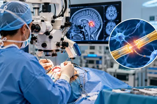
After a serious head injury, doctors first protect breathing and the cervical spine, assess consciousness and pupils, and look for bleeding or swelling on an urgent CT scan. You will learn how that information determines observation, pressure treatment or surgery, what happens in the operating room, and what families can expect during recovery and consultation.
Key takeaways
- Airway, breathing, circulation and neck protection come before brain scans.
- CT scans identify bleeding, swelling and skull fractures that can require surgery.
- Ask who will operate, why surgery is needed and what ICU risks to expect.
- Bring scan discs, reports, medication lists and the injury timeline.
What happens in the emergency department after a head injury?
Airway, breathing and circulation come first. Staff protect the cervical spine, give oxygen, support breathing, and treat low blood pressure immediately because hypoxia and poor circulation can cause secondary brain injury. They assess the Glasgow Coma Scale (GCS), then examine:
- Pupil size and reaction to light
- Movement and strength in each limb
- Seizures, vomiting and worsening drowsiness
- Blood or clear cerebrospinal fluid from the nose or ear
A single examination cannot show whether bleeding or swelling is progressing. Staff repeat the GCS, pupil checks, limb assessment and vital signs because a falling score, a newly unequal pupil or new weakness can signal dangerous pressure before the patient collapses.
Do not wait for relatives to collect documents, find old reports or seek multiple opinions. Take someone with loss of consciousness, repeated vomiting, confusion, seizure, worsening drowsiness, weakness or fluid or blood from the ear or nose to an emergency department immediately.
If the patient deteriorates, request immediate transfer to a trauma service with neurosurgical and intensive-care capability.
A search for what happens during traumatic brain for injury surgery in mumbai should start with this emergency assessment, not an outpatient consultation. The initial findings determine how quickly CT, specialist review and treatment must proceed.
Which tests show whether traumatic brain injury surgery is needed?
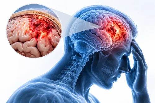
Non-contrast CT of the head is the decisive first scan for most acute traumatic brain injuries. It rapidly shows epidural haematoma, acute subdural haematoma, traumatic subarachnoid haemorrhage, contusion, hydrocephalus, midline shift, traumatic swelling and skull fracture.
Doctors add CT angiography when the injury pattern or mechanism suggests damage to a blood vessel, such as carotid or vertebral-artery dissection, pseudoaneurysm or active bleeding. MRI can reveal smaller or older injuries, but unstable patients usually undergo it later because transport, monitoring and scan time can delay urgent treatment.
Blood tests prepare the team for bleeding and anaesthesia:
- Platelet count checks the cells needed for clotting.
- PT/INR and aPTT identify clotting abnormalities or effects of blood-thinning medicines.
- Blood group and cross-match prepare compatible blood if surgery causes significant blood loss.
A scan alone does not decide the traumatic brain injury surgery process. Doctors combine its findings with GCS change, pupil abnormalities, mass effect, intracranial pressure and the patient’s overall condition.
| Approach | What it means | When it applies |
|---|---|---|
| Observation | Repeat examinations and CT scans, with medication or pressure monitoring | Stable patient without dangerous compression |
| Surgery | Evacuates blood, relieves pressure or repairs a fracture | Epidural haematoma over 30 cm³; acute subdural haematoma over 10 mm thick or with over 5 mm midline shift |
| Pressure treatment | Treats raised intracranial pressure with selected monitoring or drainage | Severe TBI with pressure above 22 mm Hg, interpreted with the examination |
A smaller clot can still require urgent surgery after GCS decline, unequal pupils or worsening mass effect.
What happens during the traumatic brain injury surgery process?
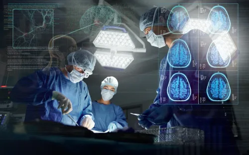
After consent, the patient receives general anaesthesia, antibiotics and blood-loss planning, including intravenous access, blood typing and blood products when needed. The team positions the body, fixes the head in a rigid holder and adds neuromonitoring when the injury or operation warrants it.
| Procedure | What happens | Purpose |
|---|---|---|
| Burr hole | A small skull opening allows selected drainage | Relieve a suitable collection |
| Craniotomy | A bone flap is removed temporarily and replaced after treatment | Access and repair the injured area |
| Decompressive craniectomy | The flap is left off to give a swollen brain room; cranioplasty replaces it later | Reduce dangerous pressure |
| Depressed-fracture elevation | Bone fragments are lifted or removed | Protect the brain and restore the skull surface |
The surgeon makes a scalp incision, opens the skull as planned, evacuates clot, controls torn vessels and treats accessible contused brain. The dura may be opened or repaired, a drain may be left, and the bone is replaced when swelling and safety permit. Staples or sutures close the scalp.
The operation prevents continuing compression and secondary injury; it cannot restore neurons destroyed by the impact. The patient then goes to intensive care for handover, where the team receives details of blood loss, medicines, drains, neurological findings and remaining concerns. Severe injury or decompressive surgery can require sedation and ventilation.
What happens after surgery in the ICU and during TBI recovery?
After surgery, a patient may remain sedated, on a ventilator or in intensive care because severe swelling, reduced consciousness, decompressive craniectomy or raised intracranial pressure makes close control safer than immediate waking. Staff repeat pupil size and reaction, limb movement and consciousness checks while controlling blood pressure, oxygen, temperature and glucose.
Seizures are treated when indicated.
- An intracranial-pressure monitor measures pressure inside the skull. A ventricular drain can also remove cerebrospinal fluid; a neurological change prompts a repeat CT.
- Nurses reposition the patient, provide chest care and assess swallowing to reduce pneumonia, blood clots and pressure injuries.
- Some patients need brief observation before leaving hospital. Others require prolonged ICU care followed by rehabilitation for walking, speech, swallowing, memory, attention, behaviour or vision.
- TBI surgery recovery in Mumbai depends heavily on the original brain injury, not only the scalp wound. Improvement can continue for months, and discharge does not mean recovery is complete.
At home, follow the wound-care instructions, give prescribed antiseizure or other medicines exactly as directed, and follow restrictions on driving and work. Keep imaging appointments and attend neurological or rehabilitation follow-up.
Seek urgent assessment for worsening headache, repeated vomiting, increasing drowsiness, seizure, new weakness, confusion, fever or wound drainage instead of waiting for a routine review.
What should relatives bring and ask at a brain injury surgery consultation in Mumbai?
Bring the original CT images on the hospital’s disc or secure digital link, not only the written report. Record the injury time and mechanism, loss of consciousness, seizures, changes in alertness, later scans, allergies, prior neurological disease and major medical history.
Bring a complete medicine list, including aspirin, clopidogrel, warfarin, apixaban and other blood thinners. Ask the treating team:
- Is emergency surgery safer than monitored observation or transfer?
- What operation is proposed, and will the bone flap be replaced?
- Will blood be needed, and what are the major risks?
- How long might ICU care, ventilation, drains or repeat imaging be required?
- What rehabilitation could be needed for movement, speech, swallowing, memory or behaviour?
| Option | What it means | Key question |
|---|---|---|
| Emergency surgery | Relieves dangerous bleeding or pressure now | What deterioration are we preventing? |
| Monitored observation | Repeat examinations and CT scans guide treatment | How often will checks and imaging occur? |
| Transfer | Moves the patient to neurosurgical or intensive-care capability | Who is responsible during transport? |
A brain injury surgery consultation in Mumbai with Dr. Anshu Warade can help relatives understand these choices and the likely recovery plan. Do not delay emergency treatment for a routine consultation if the patient has unequal pupils, worsening drowsiness, repeated vomiting, seizure, new weakness, confusion, or fluid or blood from the ear or nose.
Related services
Brain (Cranial) Surgeries 🧠 Brain (Cranial) Surgeries by Dr. Anshu Warade Brain (Cranial) Surgeries involve advanced surgical procedures to treat conditions affecting the... View service → |
Neurological Examination 🧠 Neurological Examination by Dr. Anshu Warade A Neurological Examination is a detailed clinical assessment used to evaluate the functioning of the... View service → |
Frequently asked questions
What happens in the emergency department after a head injury?
The team first secures the airway, supports breathing, treats low blood pressure and protects the cervical spine. Staff then record the Glasgow Coma Scale and perform a neurological examination.
Which tests show whether traumatic brain injury surgery is needed?
An urgent non-contrast CT head scan shows bleeding, brain swelling, skull fractures and pressure effects. Doctors combine these findings with the Glasgow Coma Scale, pupil examination and changes in neurological status before recommending surgery.
What happens during the traumatic brain injury surgery process?
After anaesthesia, the surgeon may remove a blood clot, control bleeding, repair a depressed skull fracture or remove part of the skull to relieve dangerous pressure. The exact procedure depends on the injury shown on imaging.
What should relatives bring to a brain injury surgery consultation in Mumbai?
Bring CT or MRI discs and reports, emergency records, the patient’s medication and allergy list, previous medical records, identity documents and a written timeline of the injury. Ask about the diagnosis, proposed procedure, alternatives, risks, ICU monitoring and recovery milestones.
Keywords