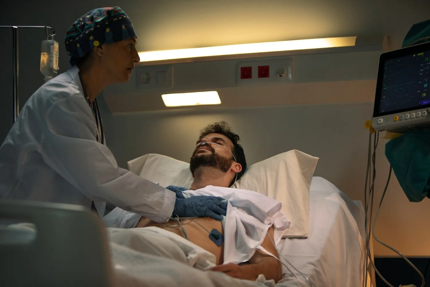
After a head injury, the first decision is not whether to book a consultation but whether the person needs emergency transport and immediate hospital assessment. You will learn which warning signs require 112 or an emergency department, how scans guide treatment, when surgery is considered, and what recovery involves after discharge.
Key takeaways
- Call 112 for loss of consciousness, repeated vomiting, seizures, or worsening drowsiness.
- Do not drive an unresponsive or confused person to an outpatient appointment.
- CT findings and neurological deterioration determine whether surgery or observation is needed.
- Recovery depends on brain injury severity, complications, rehabilitation, and follow-up care.
What to do immediately after a head injury in Worli
Call 112 immediately, or go straight to the nearest emergency department, after any head injury with loss of consciousness, worsening headache, repeated vomiting, seizure, increasing drowsiness or confusion. Do not arrange an outpatient appointment if the person is difficult to wake, has trouble walking or speaking, or is becoming less responsive.
Seek emergency assessment for any of these warning signs:
- Unequal pupils, new weakness, numbness, slurred speech or facial drooping
- Blood or clear fluid from the nose or ears
- A suspected skull fracture, a deep scalp wound or a forceful impact
- Symptoms after taking warfarin, apixaban, rivaroxaban, dabigatran, aspirin or clopidogrel
- A child or adult whose behaviour, memory or alertness worsens after appearing well
Keep the person still, support the neck, and do not give food, alcohol, sleeping tablets or aspirin. If they are vomiting, turn them carefully onto their side while protecting the neck. Do not drive someone who is confused, drowsy or having a seizure.
Whether you searched for traumatic brain injury surgery in Worli or head injury brain surgery in Mumbai, distance must not delay transfer to an appropriately equipped emergency hospital. Emergency teams can perform neurological checks and an urgent non-contrast CT; MRI is not a substitute for that first scan.
Do not give corticosteroids for traumatic brain swelling unless a specialist prescribes them for another reason.
From emergency triage to the right scan
Emergency triage starts with airway, breathing, circulation and a neurological check. Doctors record the Glasgow Coma Scale, inspect both pupils, test limb movement, and ask about loss of consciousness, vomiting, seizures, anticoagulants and the injury mechanism. A falling score, unequal pupils or new weakness requires immediate neurosurgical review, not an outpatient appointment.
The initial assessment usually includes:
- Stabilise oxygen levels, blood pressure and the neck when spinal injury is possible.
- Perform a non-contrast CT head urgently.
- Add CT cervical-spine imaging when neck pain, neurological signs, reduced consciousness or a high-risk mechanism suggests spinal injury.
- Repeat the neurological examination while awaiting imaging and monitor for deterioration.
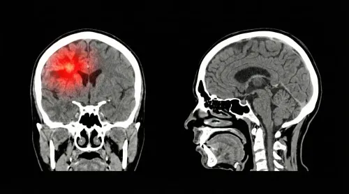
CT is the emergency decision-maker because it quickly reveals an epidural or subdural haematoma, subarachnoid blood, contusions, cerebral oedema, skull fractures and midline shift. These findings can determine whether observation, intensive-care treatment or surgery is needed.
MRI is not a faster replacement for CT. After stabilisation, doctors use it selectively for subtle diffuse axonal injury, injuries that CT does not explain, or persistent cognitive, balance or neurological symptoms.
Transfer to the nearest equipped emergency hospital should not be delayed by searching for “tbi surgery worli” or “traumatic brain injury surgeon worli”; the priority is immediate CT interpretation, neurosurgical decision-making and critical care.
How scan findings and deterioration change treatment
Doctors choose observation, repeat CT, intensive care or surgery from two moving targets: the scan and the neurological examination. A small, stable bleed with normal pupils and unchanged consciousness may be observed; falling consciousness, a new weakness, unequal pupils, repeated vomiting or a seizure demands immediate reassessment.
| Option | What it means | When it applies |
|---|---|---|
| Observation | Frequent consciousness, pupil and limb checks | Stable examination, no dangerous mass effect and a scan suitable for monitoring |
| Repeat CT | A second non-contrast head CT | Worsening symptoms, anticoagulant use, or concern that bleeding or swelling is expanding |
| Intensive care | Airway support, close neurological monitoring, seizure treatment and management of raised intracranial pressure | Reduced consciousness, breathing risk, significant swelling or a bleed needing continuous monitoring |
| Urgent surgery | Evacuation of blood and relief of pressure | Epidural or subdural haematoma causing mass effect, deterioration or dangerous pressure |
For an acute subdural haematoma, evacuation is recommended when the clot is thicker than 10 mm or midline shift exceeds 5 mm, regardless of Glasgow Coma Scale score. Smaller measurements can still require surgery when consciousness falls, pupils become abnormal or the GCS is low.
Tell the team the exact anticoagulant or antiplatelet drug, dose and last dose time. Do not stop prescribed medication independently. Routine high-dose corticosteroids are harmful in significant TBI and are not a treatment for traumatic cerebral swelling.
For traumatic brain injury surgery Worli, confirm access to emergency CT interpretation, neurosurgery, neurocritical care, repeat imaging and rehabilitation coordination. A search for brain injury recovery surgery should not delay transfer to the nearest appropriately equipped emergency hospital.
Craniotomy, craniectomy and the operation’s limits
Traumatic brain injury operations include craniotomy, craniectomy, clot evacuation and repair of open or depressed skull fractures. The choice depends on bleeding, pressure, swelling and the skull wound—not the scan alone. An epidural or subdural haematoma may need rapid evacuation; decompression addresses dangerous swelling when intensive care is insufficient.
| Operation | Bone treatment | Main purpose |
|---|---|---|
| Craniotomy | Bone flap removed and replaced | Evacuate a clot, control bleeding or repair the skull |
| Craniectomy | Bone flap removed and left out temporarily | Allow swollen brain to expand and reduce pressure |
A craniotomy removes a bone flap, permits clot evacuation or bleeding control, and replaces the flap before closing. A craniectomy leaves the flap out to give swollen brain room; after swelling settles, cranioplasty replaces it in a separate operation with infection, fluid-collection and seizure risks.
Neither operation restores brain tissue already destroyed. Recovery also depends on the initial injury, oxygen and blood-pressure control, seizures, infection, sedation, complications and rehabilitation. Routine corticosteroids do not treat traumatic cerebral swelling; high-dose methylprednisolone increased mortality in the CRASH trial.
A search for TBI surgery Worli or head injury brain surgery in Mumbai should focus on emergency CT interpretation, neurosurgical decisions, neurocritical care, repeat imaging and rehabilitation coordination—not distance alone. After serious injury, transfer to the nearest appropriately equipped emergency hospital must not wait for an outpatient appointment.
Recovery after traumatic brain injury surgery
Improvement after surgery is rarely linear. Alertness, memory, speech, balance, behaviour and fatigue can change at different speeds, with good days followed by setbacks. Intensive-care monitoring, repeat neurological examinations and follow-up CT scans help detect swelling, bleeding or hydrocephalus before it causes further damage.
Brain injury recovery surgery does not end when the wound is closed. Early rehabilitation may include physiotherapy for strength and walking, occupational therapy for daily tasks, speech-language therapy for communication and swallowing, neuropsychology for thinking and behaviour, and nursing support.
If a craniectomy left the bone flap out, ask when cranioplasty is planned; it is a separate operation after swelling settles.
A search for a traumatic brain injury surgeon Worli should lead you to check the whole pathway, not distance alone:
| Care capability | Why it matters | What to verify |
|---|---|---|
| Emergency CT interpretation | A fresh bleed or midline shift can require immediate action | Is a CT head scan available and reviewed urgently? |
| Neurosurgical and neurocritical care | Severe swelling needs airway, pressure and neurological management | Who makes decisions overnight? |
| Repeat imaging | A haematoma can enlarge after the first scan | Is repeat CT arranged when examination changes? |
| Rehabilitation coordination | Surgery cannot restore every lost function | Are physiotherapy, occupational and speech therapy referrals organised? |
Ask Dr. Anshu Warade how follow-up will cover wound checks, seizure risk, medicines and rehabilitation. Provide the exact name, dose and last-dose time for every anticoagulant or antiplatelet drug; never stop it independently. Routine corticosteroids are not standard treatment for traumatic brain swelling and can increase harm.
Related services
Brain (Cranial) Surgeries 🧠 Brain (Cranial) Surgeries by Dr. Anshu Warade Brain (Cranial) Surgeries involve advanced surgical procedures to treat conditions affecting the... View service → |
Neurological Examination 🧠 Neurological Examination by Dr. Anshu Warade A Neurological Examination is a detailed clinical assessment used to evaluate the functioning of the... View service → |
Frequently asked questions
When should you seek emergency care after a head injury in Worli?
Call 112 or go to the nearest emergency department after loss of consciousness, worsening headache, repeated vomiting, seizure, increasing drowsiness, confusion, trouble walking, trouble speaking, or reduced responsiveness.
Which scan is used to assess a suspected traumatic brain injury?
Emergency teams use neurological assessment and brain imaging to identify bleeding, swelling, skull fractures, and pressure effects. A CT scan is the key urgent scan described for this decision.
When does a traumatic brain injury require surgery?
Treatment changes when scan findings show a dangerous collection of blood, swelling, pressure, or skull injury, and when the person’s neurological condition deteriorates.
What is the difference between a craniotomy and a craniectomy?
A craniotomy removes a section of skull temporarily and replaces it after access or treatment. A craniectomy leaves the bone off temporarily to give a swollen brain more room.
What does recovery after traumatic brain injury surgery involve?
Recovery includes monitoring for complications, follow-up examinations, and rehabilitation tailored to problems with movement, speech, thinking, behaviour, or daily activities.
Keywords