The phrase “endoscopic brain surgery” covers several operations, from a procedure through the nose to an endoscope-assisted operation through a small skull opening. You will learn how surgeons investigate the problem, choose the safest route, prepare you for anaesthesia, manage the operation and monitor recovery through follow-up.
Key takeaways
- Bring symptom timelines, treatment records and changes in vision or hormone function.
- Compare endoscopic routes, open surgery and non-surgical options before choosing an operation.
- Expect checks of brain function, hormones and the surgical route during early recovery.
- Ask when pathology results, follow-up imaging and the next appointment will be available.
The consultation establishes whether surgery is appropriate
The endoscopic brain surgery consultation establishes whether surgery is appropriate, safe and likely to help. You describe your symptoms, when they began, previous treatment and any changes in vision, hormone function, balance, memory or movement.
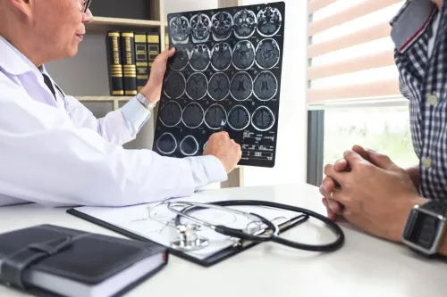
The surgeon performs a neurological examination, reviews every medicine and examines the actual MRI or CT images rather than relying only on the written report.
The team also assesses your fitness for anaesthesia. Tell them about allergies, bleeding problems, previous operations, earlier anaesthetic reactions and illnesses such as heart or lung disease.
Further tests depend on the suspected condition:
- MRI with contrast shows the lesion and its relationship to the brain and skull base.
- CT shows bone, sinus anatomy and the boundaries available for surgery.
- CT angiography or MR angiography maps arteries and venous structures near the planned route.
- Pituitary testing may measure prolactin, cortisol-related function, thyroid hormones, growth-hormone-related markers and gonadal hormones.
- Formal visual-field testing checks whether a lesion has affected the optic chiasm or optic nerves.
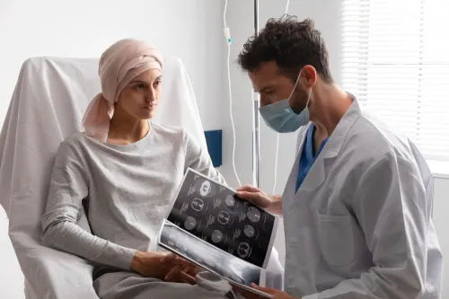
The surgeon uses these findings to identify the suspected diagnosis and explain why each test matters. The consultation should also clarify the expected benefit, the main risks and whether another treatment or observation is safer. You should leave knowing what remains uncertain and what decision the next test will inform.
The surgeon compares routes and alternatives before choosing an operation
Endoscopic brain surgery is an umbrella term, not one standard operation. The route depends on the lesion’s location, size, pathology and relationship to the optic nerves, carotid arteries, other vessels and cerebrospinal-fluid pathways. The endoscopic brain surgery process therefore starts with choosing access, not with assuming that a smaller opening is safer.
| Option | Access or treatment | When it applies |
|---|---|---|
| Endoscopic endonasal | Through one or both nostrils, nasal passages and sphenoid sinus | Selected pituitary and skull-base conditions |
| Endoscope-assisted keyhole | Through a small scalp incision and skull opening | Targets reachable through a limited cranial corridor |
| Endoscopic ventricular procedure | Through a small opening into the brain’s fluid spaces | Conditions such as hydrocephalus treated by endoscopic third ventriculostomy |
| Open craniotomy | A larger skull opening | Large, distant or tightly attached lesions when it provides safer control |
| Stereotactic treatment, radiation, medication or observation | Non-open treatment or surveillance | Lesions suited to focused treatment, medical control or monitoring |
Ask how each option affects tumour control, hormone levels, vision, recovery time, repeat-treatment risk and complications. What happens during endoscopic brain surgery is shaped by these trade-offs, including the possibility of cerebrospinal-fluid leakage, bleeding or injury to critical structures.
Complete removal is not always the safest endpoint. If tissue is inseparable from the carotid artery, optic apparatus or cavernous sinus, planned residual disease followed by surveillance, medication or radiotherapy can reduce harm. Dr. Anshu Warade can help patients compare these route-specific choices during treatment planning.
Related services
Endoscopic Brain Surgery 🧠 Endoscopic Brain Surgery by Dr. Anshu Warade Endoscopic Brain Surgery is an advanced, minimally invasive neurosurgical technique that uses a thin... View service → |
Skull Base Neurosurgery 🧠 Skull Base Neurosurgery by Dr. Anshu Warade Skull Base Neurosurgery is a highly specialized field that focuses on treating complex tumors and... View service → |
Preparation and the operating-room sequence happen in defined steps
Before admission, the hospital gives you instructions on fasting, permitted medicines, bathing and infection screening. Do not stop aspirin, clopidogrel, warfarin or a direct oral anticoagulant unless the surgical or prescribing team gives explicit instructions.
1. The team makes a blood-thinner plan and completes an anaesthesia assessment. This checks your airway, heart, lungs, medication list and previous anaesthetic reactions. Arrange an adult to take you home and support you after discharge.
2. You sign consent after discussing the planned route, the possibility of changing to an open operation, blood transfusion, residual disease and likely complications. This is part of understanding the endoscopic brain surgery process, not a formality.
3. On the day, staff confirm your identity, allergies, consent, procedure and scan review before placing an intravenous line. The procedure uses general anaesthesia when required. The team positions you for safe access and stabilises your head when necessary.
4. For skull-base surgery, image guidance can match preoperative CT or MRI data to your anatomy and orient the team around the orbit, carotid arteries, optic nerves and skull-base boundaries. It supports judgement; it does not remove injury risk.
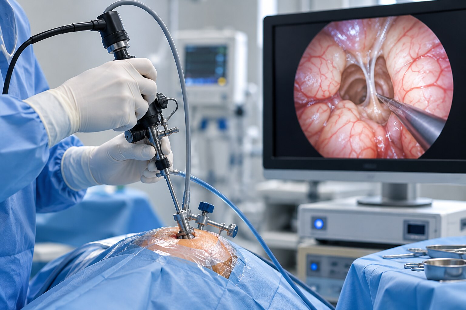
In an endonasal operation, the surgeon passes a rigid endoscope and instruments through one or both nostrils, opens the nasal and sphenoid pathways in stages, reaches the target, then closes any cerebrospinal-fluid opening with graft material or a vascularized nasoseptal flap when needed.
Early recovery focuses on the brain, hormones and the surgical route
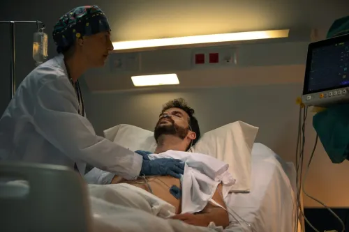
Within the first hours after surgery, nurses and doctors check consciousness, strength, vision, pain, nausea and risks specific to the operation. Pituitary and skull-base patients also need urine-volume records, fluid-balance checks, serum sodium tests and pituitary hormone assessment. Diabetes insipidus, low sodium or adrenal insufficiency can require immediate treatment or repeat blood tests after discharge.
| Procedure | Typical recovery pattern | What can lengthen the stay |
|---|---|---|
| Small ventricular procedure | Often a shorter admission and quicker return to basic activity | Blocked drainage, bleeding or neurological change |
| Endonasal pituitary or skull-base surgery | Brain monitoring continues while nasal healing progresses | CSF leak, hormone disturbance, visual problem or major blood loss |
| Extensive skull-base tumour operation | More prolonged observation and rehabilitation needs | Vascular injury, significant bleeding or complex reconstruction |
Nasal healing is separate from brain recovery. Congestion, bloody drainage, reduced smell and crusting are common after an endonasal route; saline irrigation, nasal endoscopy or debridement may be prescribed. Do not blow your nose, strain or lift until the team permits it, especially after a cerebrospinal-fluid repair.
Before leaving, confirm:
- Which medicines to take and when
- When you may shower
- How to care for the nose or wound
- Activity limits and warning signs
- The date of your first review
Endoscopic brain surgery recovery is measured by safe function, stable vision and hormone control, not simply by the absence of a scalp incision.
Pathology, imaging and follow-up determine what happens next
Pathology, postoperative imaging and follow-up determine what happens next. After discharge, pathology identifies the tissue type when a specimen is removed. A postoperative MRI or CT shows residual disease, the repair and the new baseline.
For pituitary disease, hormone levels and formal visual-field testing show whether treatment corrected hormone excess or relieved pressure on the optic pathways.
What happens during endoscopic brain surgery is only part of the decision. Follow-up may involve surveillance MRI, endocrine testing and nasal examinations, or lead to medication, repeat surgery or radiation when residual or recurrent disease needs treatment.
Ask when each test is due, which clinician will interpret it, whether the intended endpoint was achieved, whether any lesion remains, which restrictions still apply and how recurrence will be detected. The final assessment includes safe healing, preserved vision and hormone function, a confirmed diagnosis and a plan for the next stage of care.
Pathology and imaging commonly lead to one of these plans:
| Option | What it means | When it applies |
|---|---|---|
| Surveillance | Repeat MRI, hormone tests or nasal examinations | No treatment is needed for stable or fully treated disease |
| Medication | Drug treatment for hormone activity or residual disease | Hormone excess or controllable remaining tissue persists |
| Repeat surgery | A second operation removes or relieves remaining disease | Residual or recurrent disease is accessible and treatment is worthwhile |
| Radiation | Targeted treatment controls remaining or returning tissue | Surgery or medication cannot safely achieve control |
Seek urgent advice for:
- Persistent clear, watery nasal drainage
- Fever, worsening headache or neck stiffness
- New visual loss or confusion
- Marked thirst with very frequent urination
- Inability to keep fluids down
Frequently asked questions
What happens during an endoscopic brain surgery consultation?
You discuss your symptoms, their timing, previous treatment and changes in vision, hormone function, balance, memory or movement. The surgeon reviews examinations and scans to decide whether surgery is appropriate, safe and likely to help.
How does the surgeon choose the surgical route?
The surgeon compares the target’s location, its relationship to the brain and nearby structures, the risks of each route and alternatives such as observation, medication or open surgery.
What happens during the endoscopic brain surgery process?
Preparation includes medical assessment, scan review and surgical planning. In the operating room, the team administers anaesthesia, positions you, reaches the target through the planned route, treats or removes the lesion, and closes the access point.
What is endoscopic brain surgery recovery like?
Early recovery includes monitoring brain function, vision, hormone levels and symptoms related to the surgical route. Your care team also checks pain, nausea, movement and wound healing before discharge planning.
What happens after pathology and follow-up imaging?
Pathology identifies the tissue diagnosis when tissue was removed. Follow-up imaging shows the treatment result, and your surgeon uses both findings to plan surveillance, additional treatment or routine recovery care.