A safe cranial operation starts long before the first incision: the surgeon must confirm the diagnosis, define the purpose of treatment and compare surgery with reasonable alternatives. By the end, you will know which records to bring, which planning decisions matter and how to compare a surgeon’s experience, hospital support and proposed care plan.
Key takeaways
- Bring symptom timelines, medication lists, scans, and questions to your consultation.
- Ask what non-surgical options exist and why surgery offers greater benefit.
- Compare the surgeon’s experience, hospital support, risks, and recovery plan.
- Confirm who manages your care before admission, after discharge, and during rehabilitation.
What should happen during the first brain doctor consultation?
A best brain doctor consultation turns your story into one focused clinical question, not simply “Can this lesion be removed?” Explain when symptoms began, how they changed, and whether you have headaches, seizures, weakness, speech or vision changes, memory problems, or reduced ability to work, walk, drive, care for yourself or manage daily tasks.
Describe previous treatments and their results.
Bring:
- MRI or CT images on a disc or portal link
- Radiology reports and pathology reports
- Blood-test results
- A complete medication list with doses
- Prior operative notes
The surgeon will review every medicine because it can change surgical safety and recovery. Include anticoagulants such as warfarin or apixaban, antiplatelet drugs such as aspirin or clopidogrel, steroids, antiseizure medicines, diabetes medicines and supplements. Do not stop a blood thinner or antiseizure medicine without an agreed plan from the surgical team and prescriber.
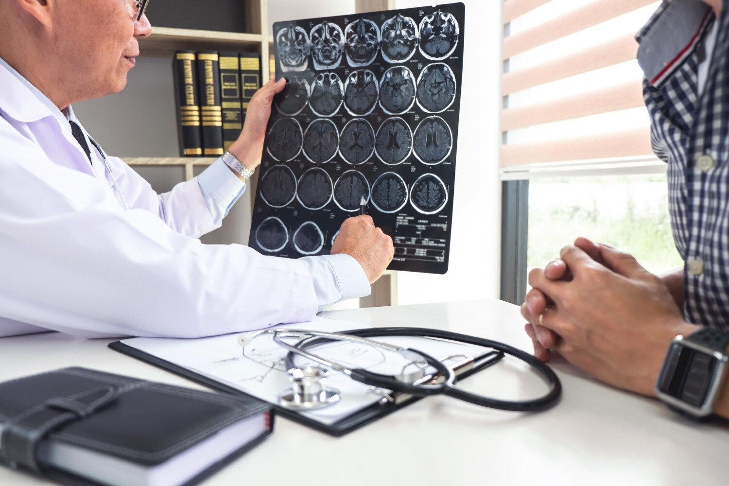
A neurological examination tests strength, sensation, coordination, reflexes, speech, vision, eye movements and memory; it adds functional information that a scan report cannot provide. The consultation should finish with a clear goal: removal, decompression, tissue diagnosis, seizure control, symptom relief, or preservation of speech, movement or vision. Ask: What is the diagnosis?
How urgent is treatment? What benefit should I expect? What are the risks of waiting? Do I need another specialist review?
How does the surgeon decide whether cranial surgery is the right treatment?
An abnormal scan does not automatically mean an operation. A brain doctor for surgery compares symptoms, neurological examination, MRI with contrast and specialized sequences, CT findings, growth on serial scans, and overall health. A stable finding without symptoms may be observed; worsening weakness, seizures or pressure can make treatment urgent.
CT is particularly useful for urgent bleeding, calcification, skull injury and bone anatomy. MRI usually gives more detail about brain tissue and lesions, helping define their boundaries and relationship to critical structures.
The alternatives may include observation with repeat imaging, medication, needle or surgical biopsy, radiation, stereotactic radiosurgery, endovascular treatment or a second opinion. The diagnosis and goal determine the route:
- A tumor plan may prioritise maximal safe removal and tissue for histology and molecular classification.
- Hydrocephalus may require temporary drainage or shunt planning.
- An aneurysm may lead to clipping or endovascular treatment.
- Trauma may require urgent evacuation of blood or pressure control.
- Epilepsy surgery requires seizure localization before removing or disconnecting tissue.
- A skull-base lesion, or one near language, motor or visual pathways, demands function-preservation planning.
Cranial surgery planning must compare the risk of operating with the risk of not operating. Consent should cover bleeding, infection, seizures, stroke, swelling, cerebrospinal-fluid leak, new neurological deficit, further surgery and stopping resection to preserve function, alongside the expected benefit of each option.
Which technical decisions shape the cranial surgery plan?
A detailed cranial surgery planning document states the surgical route, craniotomy size and location, patient position, anaesthetic plan, monitoring, and whether tissue is needed for diagnosis. The route must reach the lesion while protecting nearby language, motor, sensory, visual and vascular structures.
| Approach | Purpose | Main consideration |
|---|---|---|
| Open craniotomy | Expose the lesion for removal or biopsy | More exposure, with greater disruption than a keyhole route |
| Minimally invasive access | Reach selected lesions through a smaller opening or corridor | Suitable only when the target and trajectory are safe |
| Needle or limited biopsy | Obtain tissue for diagnosis | Provides less treatment and may miss mixed tumour areas |
General anaesthesia is common; selected lesions near critical networks require an awake craniotomy so the patient can perform speech or movement tasks during mapping. Positioning is chosen for a safe line of sight, stable access and protection of pressure points.
Functional MRI, diffusion-tensor tractography and neuropsychological testing help predict language, memory and movement risks. Functional MRI and tractography guide planning but do not replace direct cortical stimulation when speech or movement is at risk.
Neuronavigation matches instruments to preoperative scans, but brain shift after skull opening, cerebrospinal-fluid release or tissue removal reduces accuracy. Intraoperative MRI, ultrasound or updated navigation imaging can correct that shift. Neurophysiological monitoring tracks threatened function, and the surgeon may stop resection rather than cause a permanent deficit.
Complex cases need review by neurosurgery, neuroradiology, neuropathology, neuro-oncology, radiation oncology, neurology, neuroanaesthesia and rehabilitation.
How do you assess a brain doctor and hospital for a specific operation?
Judge a surgeon by the operation you need, not by the label “best brain doctor for surgery.” Confirm appropriate board certification, regular performance of the proposed operation, and experience with your exact condition, surgical approach and risk profile.
Ask what the hospital can provide for that procedure:
- Neuroanesthesia and a neurosurgical intensive care unit
- Neurophysiological monitoring and intraoperative imaging
- Neuropathology and blood-bank support
- Rehabilitation, emergency coverage and overnight specialist response
- Named infection-prevention measures for the operating room and ward
| Question to ask | Useful answer | Warning sign |
|---|---|---|
| How will speech or movement be protected? | Mapping, monitoring and a clear stopping threshold | “We will know during surgery” without detail |
| Is tissue diagnosis required? | A reasoned plan for biopsy, resection or no tissue sampling | No explanation of how diagnosis changes treatment |
| What complication rate applies in your practice? | Procedure-specific figures and definitions | Only hospital-wide or promotional numbers |
| What if complete removal is unsafe? | A plan for residual disease, radiation, further surgery or rehabilitation | No contingency plan |
A consultation with Dr. Anshu Warade can help assess the diagnosis, operative indication, alternatives and hospital pathway rather than relying on general reputation. Seek a second opinion when the diagnosis is uncertain, urgency is unexplained, or the operation carries major functional risk.
Related services
Brain (Cranial) Surgeries 🧠 Brain (Cranial) Surgeries by Dr. Anshu Warade Brain (Cranial) Surgeries involve advanced surgical procedures to treat conditions affecting the... View service → |
Skull Base Neurosurgery 🧠 Skull Base Neurosurgery by Dr. Anshu Warade Skull Base Neurosurgery is a highly specialized field that focuses on treating complex tumors and... View service → |
How does planning continue from admission through recovery?
Cranial surgery planning continues after admission because medication safety, early imaging and functional recovery determine whether discharge is safe. A best brain doctor consultation should leave you with a written plan for the days before and after surgery.
1. Confirm instructions for anticoagulants, antiplatelet medicines, diabetes treatment, steroids and antiseizure drugs before admission. Never stop a prescribed blood thinner or antiseizure medicine without an agreed plan from the surgical team and the relevant prescriber.
2. Steroids treat brain edema or mass effect when needed, then the dose is reduced as clinically appropriate. Prolonged use can cause high blood glucose, infection risk, mood or sleep changes and muscle weakness. Antiseizure treatment is individualized; it is not automatically continued forever after every craniotomy.
3. An immediate postoperative scan checks for bleeding and other complications and establishes how much target tissue remains. After glioma surgery, MRI is commonly obtained within 24–48 hours because later treatment-related enhancement can obscure residual disease.
4. Recovery assessment covers swallowing, speech, cognition, strength, mobility, pain, wound condition and home support. Speech, occupational, physical or cognitive rehabilitation is arranged when those functions are affected. Discharge depends on neurologic function and reliable support, not the incision alone.
Frequently asked questions
What should happen during the first brain doctor consultation?
The consultation should turn your symptoms, examination findings, medical history, and scan results into a clear clinical question. Describe when symptoms began, how they changed, and how they affect work, mobility, speech, vision, memory, driving, and daily tasks. Bring scan discs and reports, medication details, previous treatment records, and a written question list.
How does a surgeon decide whether cranial surgery is the right treatment?
The surgeon weighs the diagnosis, symptoms, lesion location, growth or progression, expected benefit, surgical risks, and alternatives such as monitoring, medication, radiation, or further testing. Ask what happens if you delay treatment and which finding would change the recommendation.
Which technical decisions shape a cranial surgery plan?
Planning includes the surgical route, the patient’s position, how much tissue to remove or treat, protection of speech and movement areas, imaging or navigation tools, anaesthesia needs, blood-loss planning, and access to intensive care. The correct choices depend on the condition, scan findings, and the brain structures involved.
How do you assess a brain doctor and hospital for a specific operation?
Ask whether the surgeon regularly performs your operation, who provides anaesthesia and critical care, which specialists support the case, and how complications are handled. Check the hospital’s imaging, intensive-care, rehabilitation, infection-control, and emergency arrangements for the planned procedure.
How does planning continue from admission through recovery?
Before admission, confirm medications, fasting instructions, tests, arrival time, consent, and post-operative support. After surgery, ask how neurological checks, pain control, wound care, scan reviews, discharge decisions, rehabilitation, driving, work, and follow-up appointments will be managed.
Related services
Endoscopic Brain Surgery 🧠 Endoscopic Brain Surgery by Dr. Anshu Warade Endoscopic Brain Surgery is an advanced, minimally invasive neurosurgical technique that uses a thin... View service → |
Neurological Examination 🧠 Neurological Examination by Dr. Anshu Warade A Neurological Examination is a detailed clinical assessment used to evaluate the functioning of the... View service → |